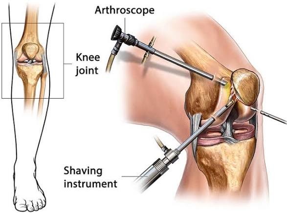
Arthroscopy is a minimally invasive "keyhole" surgery that uses a small camera (arthroscope) and tiny instruments inserted through small cuts to diagnose and treat joint problems in areas such as the knees, shoulders, hips, ankles, elbows, and wrists. It offers less pain and faster recovery than traditional open surgery by allowing surgeons to view and repair joint damage.
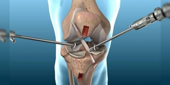
The procedure involves inserting the arthroscope to visualize the joint on a screen, followed by the use of specialized instruments to trim cartilage, repair ligaments (such as ACL), or address other joint issues. It is commonly performed under general, spinal, or local anesthesia.
What It’s Used For
- Diagnosing persistent joint pain, swelling, stiffness, or instability not visible on X-rays.
- Treating conditions such as torn meniscus, ACL injuries, rotator cuff tears, and some forms of arthritis.
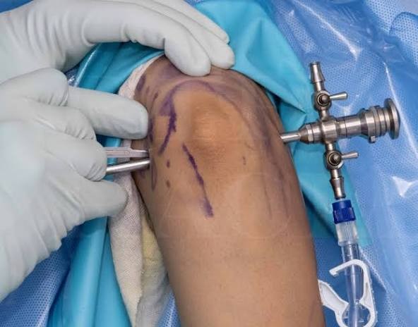
The Procedure
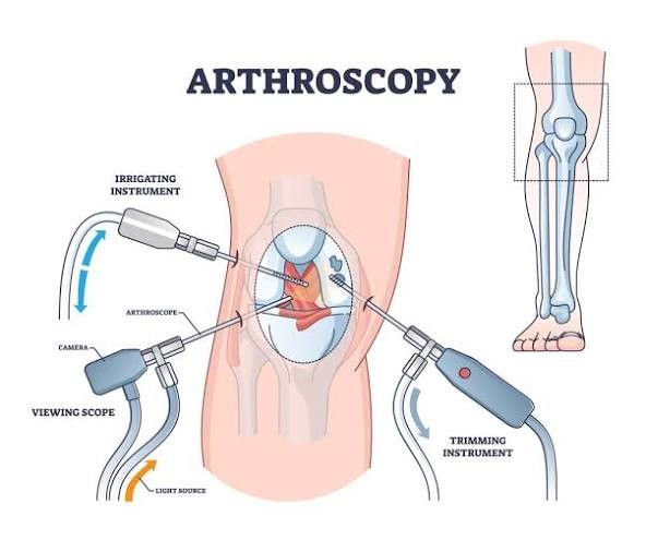
- Anesthesia: General (patient asleep), spinal (lower body numb), or local (specific area numb).
- Incisions: Small cuts (2–3 mm) are made near the affected joint.
- Arthroscope: A thin tube with a camera and light is inserted to view the joint.
- Treatment: The surgeon uses the video feed to guide instruments and repair damage (e.g., trimming cartilage).
- Closure: Incisions are closed using tape or stitches.
Benefits
- Smaller incisions with less pain and reduced scarring.
- Lower risk of infection and bleeding compared to open surgery.
- Faster recovery and earlier return to daily activities.
Related Images